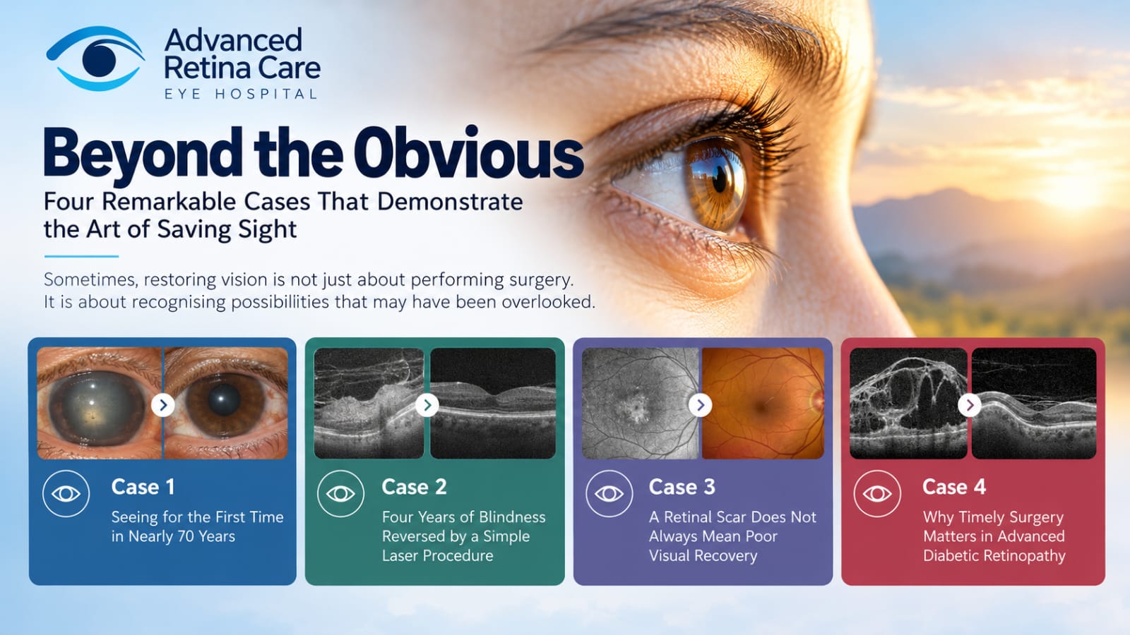
Sometimes, restoring vision is not just about performing surgery. It is about recognising possibilities that may have been overlooked. At Advanced Retina Care, we believe that every eye deserves a careful assessment, particularly when a patient has been told that nothing more can be done to improve their vision.
Over the years, I have encountered several patients whose visual prognosis was initially considered extremely poor. Some had lived with visual impairment for decades, while others had undergone previous retinal surgeries or had been told that their retinal condition made visual recovery impossible.
These four cases illustrate an important principle of retinal care: never underestimate the importance of a detailed examination, sound clinical judgement and identifying potentially reversible causes of visual loss.
- Case 1: Seeing for the First Time in Nearly 70 Years
- Case 2: Four Years of Blindness Reversed by a Simple Laser Procedure
- Case 3: A Retinal Scar Does Not Always Mean Poor Visual Recovery
- Case 4: Why Timely Surgery Matters in Advanced Diabetic Retinopathy
- What These Four Cases Teach Us (Five Core Principles)
- Our Philosophy at Advanced Retina Care
Case 1: Seeing for the First Time in Nearly 70 Years
One of the most memorable patients I encountered was a woman in her seventies who had apparently been blind in one eye since birth.
She had a very large squint and a dense, black cataract. Over several decades, she had consulted multiple doctors, but no one had attempted to restore vision in that eye. The longstanding blindness and obvious squint had led to very limited expectations of visual recovery.
She initially came to us because of diabetic retinopathy in her other eye.
The Critical Clinical Finding
During examination, I noticed that the intraocular pressure in the apparently blind eye was completely normal. High-resolution imaging suggested peripheral tractional retinal detachment, but importantly, the macula appeared to be spared.
I felt that the eye might still have useful visual potential and decided to attempt cataract surgery after counselling the patient.
The result was extraordinary.
On the first postoperative day, her vision was 20/100. She became emotional because she could see through that eye for the first time in approximately seventy years.
I subsequently performed surgery for the tractional retinal detachment. Her vision improved further to 20/50. Despite the longstanding squint, she did not experience double vision.
Clinical Takeaway
An eye that appears to have no visual potential may sometimes still have a chance of meaningful visual recovery. Never assume longstanding blindness with squint precludes useful sight without an anatomical macular evaluation.
Case 2: Four Years of Blindness Reversed by a Simple Laser Procedure
A gentleman of approximately 65 years came to my clinic with severe visual impairment in both eyes. He had previously undergone vitrectomy surgery for diabetic tractional retinal detachment.
His vision was:
- Right eye: No perception of light (NPL).
- Left eye: Hand movements only.
The right eye had significant optic atrophy. The left eye also had a pale-looking optic disc, and the retina was flat following previous surgery.
The Clinical Discrepancy
Something did not quite match. The degree of optic atrophy in the left eye did not appear sufficient to explain the profound loss of vision. On further high-magnification examination, I suspected that posterior capsular opacification (PCO) was contributing significantly to his poor vision.
I recommended an Nd:YAG laser posterior capsulotomy.
Initially, the patient was reluctant. He had lived with poor vision for approximately four years and had understandably lost confidence that anything could improve his sight.
After counselling him about the procedure, he agreed. The result was remarkable.
His left-eye vision improved from hand movements to 20/40. The patient and his family were overwhelmed with emotion.
Clinical Takeaway
We must not automatically attribute all visual loss following retinal surgery to optic atrophy. Even when permanent retinal or optic nerve damage exists, there may be an additional, treatable cause of poor vision hiding in plain sight.
Case 3: A Retinal Scar Does Not Always Mean Poor Visual Recovery
A lady recently came for review following cataract surgery in her right eye.
Before surgery, her vision was 20/60. She also had a small area of macular atrophy. She had previously been told that her vision would not improve because of the retinal abnormality.
High-Resolution Macular Mapping
When I examined her macula, I noticed that the area of atrophy did not involve the centre of the fovea. This was an important observation. The presence of macular atrophy does not necessarily mean that cataract surgery cannot improve vision. The precise location of the retinal damage, especially its relationship to the foveal centre, is extremely important.
After explaining the likely visual potential, she decided to proceed with cataract surgery.
Her postoperative vision improved to 20/30, with N6 near vision.
Clinical Takeaway
Carefully assess the macula with OCT and dilated biomicroscopy rather than assuming that every retinal abnormality or scar will prevent visual improvement following cataract surgery.
Case 4: Why Timely Surgery Matters in Advanced Diabetic Retinopathy
Another patient presented with very severe proliferative diabetic retinopathy.
She had already undergone retinal laser treatment and received intravitreal anti-VEGF injections. However, I noticed increasing traction along the retinal arcades.
I was concerned about progressive tractional retinal detachment and advised prompt vitreoretinal surgery.
Unfortunately, the patient declined surgery at that time. Approximately three months later, she underwent surgery elsewhere. When she subsequently came back to see me, she had profound visual loss in that eye.
By then, the retinal condition appeared to have progressed considerably, with very little visual potential remaining.
Why Delay Proves Costly in Retinal Traction
This was a particularly difficult case because I had recognised the increasing traction earlier and had advised timely surgery. It illustrates an important message for patients with advanced diabetic retinopathy: progressive retinal traction should never be ignored. Regular retinal examinations, appropriate treatment and timely surgical intervention can be crucial in preserving vision.
What These Four Cases Teach Us
Every patient is different, and no two retinal conditions behave in exactly the same way. However, these experiences have reinforced several core principles that guide my approach to vitreoretinal care at Advanced Retina Care:
Look Beyond Appearances
A longstanding history of poor vision does not always mean that visual recovery is impossible. Never rely on assumptions without anatomical retinal confirmation.
Identify Reversible Causes
Not every instance of severe visual impairment is entirely due to permanent retinal or optic nerve damage. Secondary reversible factors (like PCO or cataract) may be treatable.
Understand the Macula
The exact location of retinal damage—specifically whether the central fovea is anatomically spared—makes a significant difference when predicting visual outcomes.
Recognise Progressive Disease
Advanced diabetic retinopathy and increasing retinal traction require timely assessment and treatment. A surgical window exists where vision can be saved before traction becomes irreversible.
Individualise Every Decision
The best treatment depends on the patient's specific retinal condition, visual potential and overall clinical findings, tailored with compassionate, honest counseling.
Our Philosophy at Advanced Retina Care
At Advanced Retina Care Eye Hospital, Hyderabad, our approach is centred on careful examination, individualised treatment planning and doing everything medically appropriate to preserve or restore useful vision.
We believe that patients deserve a thorough explanation of their condition, an honest assessment of their visual potential and consideration of all appropriate treatment options.
Sometimes, the difference between poor vision and meaningful visual recovery lies in recognising one small but important clinical finding.