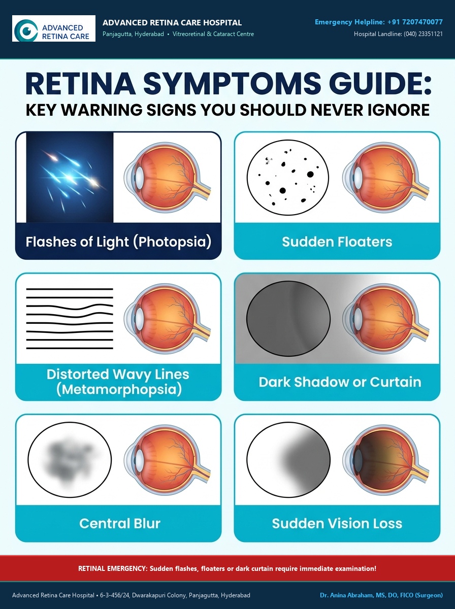
The retina is the light-sensitive layer at the back of the eye that is responsible for converting light into signals that allow us to see. Retinal problems can develop gradually, but some retinal conditions can cause sudden changes in vision that require urgent attention.
Many patients notice a change in their eyesight but are unsure whether it is serious. Symptoms such as flashes, floaters, sudden blurring, distortion, missing areas of vision or sudden loss of sight can sometimes be important warning signs of retinal disease.
This guide explains the common symptoms associated with retina problems, what they may mean, and when you should seek an examination by a retina specialist.
Comprehensive Retinal Warning Signs Guide
1. Flashes and Floaters
One of the most common symptoms that can bring a patient to a retina specialist is the sudden appearance of flashes or floaters.
If you experience flashes in eyes, you may notice brief streaks or bursts of light even when there is no obvious source of light around you. Patients may describe flashes of light in eye as small sparks, lightning-like streaks or brief flashes appearing at the edge of their vision. Some people notice flashing lights in vision, particularly when moving their eyes or looking at a dark background.
If you are seeing flashes in one eye, especially when this is a new symptom, it is important to have that eye examined because flashes can sometimes occur when the vitreous gel is pulling on the retina. The sudden appearance of flashes in eye should not be ignored, particularly if accompanied by new floaters or a shadow in your vision. Some patients notice eye flashes at night because flashes can be easier to see against a dark background. People sometimes describe seeing stars in vision, which may appear as tiny points or sparkles of light; the clinical significance depends on when they occur and whether they are associated with other visual symptoms.
Understanding Floaters, Spots & Moving Shadows
A sudden floater in eye may appear as a dark dot, thread, ring or small shape moving across your field of vision. If you suddenly notice black spots in vision, particularly if there are many new spots, you should arrange a retinal examination promptly.
Common Patient Descriptions of Floaters:
- Black dots floating in vision: Moving with eye motion and drifting across the sight.
- Spots floating in eyes: Darting away when you try to look directly at them.
- Something floating in my eye: Sudden, unexpected appearance of debris in the visual field.
- Moving spots in vision: Shadow casts from condensed collagen fibers within the vitreous onto the retina.
- Cobwebs in vision: Fine strands, lace-like or web-like structures drifting across your sight.
- Thread-like things or lines in vision: Stringy vitreous condensations moving with gaze shifts.
- Shadows floating in vision: Especially noticeable against a bright wall, white computer screen, or clear blue sky.
If you develop new floaters in eye, especially when they appear suddenly, a retina examination is required to rule out a retinal tear. A sudden increase in eye floaters is particularly important because it can occur when there is an acute posterior vitreous detachment (PVD) or when a retinal tear has ruptured a microscopic blood vessel.
When flashes and new floaters occur together, patients describe the condition as flashes and floaters. If you suddenly develop flashes and floaters in one eye, you should have that eye examined promptly because these symptoms can indicate an impending retinal detachment.
When Are Flashes and Floaters an Emergency?
Flashes and floaters are not always dangerous—they can occur as part of normal age-related liquefaction of the vitreous. However, a sudden onset or sudden shower of symptoms requires urgent examination because retinal tears can develop without causing any physical pain.
If flashes or floaters are accompanied by a curtain, dark shadow, or loss of peripheral vision, seek urgent retinal evaluation within 24 hours.
2. Sudden Blurred Vision
Sudden changes in clarity of vision can have many causes, and the timing and nature of the change are important clinical clues.
If you experience sudden blurred vision, particularly if it develops over minutes or hours, you should not simply assume that you need a new pair of glasses. Some patients describe sudden blurry vision as though a transparent film or dense fog has suddenly appeared over their eye.
If there is sudden blurred vision in one eye, comparing each eye separately by covering one eye at a time can help you recognise that the problem is isolated to one side. Patients may describe sudden blurry vision in one eye as a sudden loss of sharpness while the other eye continues to see normally.
Clinical Evaluation: When someone says their vision is suddenly blurry, it is essential to establish whether the change was genuinely sudden or whether vision had been gradually deteriorating unnoticed. A patient may notice a sudden loss of clarity in one eye when reading, looking at a face, or viewing distant street signs.
Blurry vision in one eye can occur for many reasons, including conditions involving the retina, macula, cornea, lens, or optic nerve. If you notice blurred vision in right eye or blurred vision in left eye, an examination by a retinal specialist can determine whether the cause is retinal swelling (macular edema), vascular occlusion (vein blockage), bleeding, or inflammation.
If your eyesight is suddenly getting worse over a short period, it should never automatically be attributed to ageing. If you are asking, "Why is my vision suddenly blurry?", a dilated retinal examination is the most definitive way to determine whether the problem is related to the retina or another part of the eye.
3. Sudden Vision Loss (Painless Ocular Emergency)
Sudden Vision Loss Requires Immediate Medical Care
Sudden loss of vision is a symptom that should always be taken with the highest level of urgency. If you experience sudden loss of vision in one eye, you should seek immediate ophthalmic assessment because several causes of sudden visual loss are time-sensitive emergencies.
Patients may describe this experience by saying:
- "I experienced sudden vision loss in one eye."
- "There was a sudden loss of eyesight in one eye without any warning."
- "I went blind in one eye suddenly."
- "My vision gone in one eye completely."
- "I can't see out of one eye."
- "I became suddenly blind in one eye."
Sudden painless vision loss can be caused by several serious conditions, including:
An eye stroke requiring immediate evaluation within hours.
Severe vein blockage causing widespread hemorrhage & swelling.
Sudden bleed from diabetic vessels or retinal tear filling the eye cavity.
The neurosensory retina peeling off the back of the eye.
If you lose vision suddenly, do not wait overnight hoping it will improve. Immediate evaluation at a specialized eye hospital like Advanced Retina Care can be crucial in saving your sight.
4. Curtain, Shadow or Missing Vision
A symptom that strongly suggests a retinal problem is the perception of a shadow or curtain advancing across the field of vision.
Patients commonly describe this as:
- A curtain in vision or curtain over vision
- A shadow in vision or shadow over vision
- A black shadow in eye or dark shadow in eye
- A sensation of a dark shade or blind being pulled across one eye
This symptom often begins in the peripheral vision—either at the top, bottom, or side—and progressively spreads toward the center. Some patients describe it as "half vision gone" or feeling that a section of their visual field has disappeared completely.
Other patients notice a loss of peripheral vision, a blind spot in vision, or a dark spot in vision that stays fixed in one part of their sight. If you notice that you are missing objects on one side, bumping into doorframes, or feeling like your vision has narrowed into tunnel vision, this should be investigated.
A dark curtain or shadow that spreads across your vision is one of the classic signs of a retinal detachment. This happens because the detached portion of the retina is no longer receiving nutrients and cannot transmit visual signals, creating a black or grey shadow in the corresponding field of view.
5. Distorted Vision & Wavy Lines (Metamorphopsia)
When the macula—the tiny central portion of the retina responsible for fine, detailed vision—is affected, images can become distorted. This condition is medically termed metamorphopsia.
Patients often describe this symptom by saying:
- "Straight lines look wavy."
- "There are wavy lines in vision when I read."
- "Lines look curved or bent."
- "Door frames look curved or window blinds appear bent."
- "Faces look distorted, warped or uneven."
- "Text looks irregular, with letters appearing larger or smaller than normal."
Why Do Straight Lines Look Wavy?
Straight lines look wavy when the normally flat layer of photoreceptors in the macula is physically elevated, stretched, or wrinkled by fluid, abnormal blood vessels, or scar tissue beneath or above the macula. Common causes include wet age-related macular degeneration (Wet AMD), macular edema (swelling) from diabetes, central serous chorioretinopathy (CSCR), or an epiretinal membrane (macular pucker).
6. Central Vision & Reading Problems
The macula provides the sharp, color-sensitive central vision required for reading, recognizing faces, driving, and seeing fine details.
Patients with central retinal problems may notice:
- A dark spot in center of vision or a black spot in center of eye
- A blind spot in center of vision or a gray smudge right where they are looking
- A dark patch in vision that blocks the center of a word or sentence
- Difficulty seeing faces clearly: You can see someone's hair and ears, but their central facial features are missing or blurred
- Difficulty reading: Letters disappearing or requiring significantly brighter light to decipher text
If you notice a persistent dark or distorted patch in the center of your sight, an Optical Coherence Tomography (OCT) scan of the macula is essential to diagnose conditions such as macular degeneration, macular holes, or diabetic macular swelling.
7. Diabetes & Vision Symptoms
Diabetes is one of the most important systemic causes of retinal disease. Diabetic retinopathy can damage retinal blood vessels silently over many years before visual symptoms emerge.
Patients with diabetes may experience:
- Fluctuating vision: Eyesight that changes from day to day or hour to hour with blood glucose shifts
- Gradual blurring: Caused by diabetic macular edema (leakage of fluid into the center of the retina)
- Sudden shower of dark floaters or dark streaks: Caused by fragile new blood vessels bleeding into the vitreous gel
- Sudden loss of vision: Resulting from vitreous hemorrhage or tractional retinal detachment
Diabetic retinopathy can cause significant internal damage to your retina long before you notice any blurriness or discomfort. Every person with diabetes should undergo a comprehensive dilated retinal examination at least once a year, even if their vision feels completely normal.
8. Age-Related Vision Changes
As we grow older, changes occur in the eye that can affect both the front and back structures.
Many patients assume that worsening eyesight is simply an inevitable part of getting older. While mild changes in near vision (presbyopia) and gradual clouding of the lens (cataract) are common with age, significant visual impairment, sudden blurring, distortion, or blind patches are not normal ageing.
Conditions such as age-related macular degeneration (AMD), vascular blockages, and vitreous changes become more frequent with advancing years. An examination by an eye specialist can distinguish between common age-related conditions like cataracts and serious retinal diseases that require specialized vitreoretinal treatment.
9. Symptoms Affecting Only One Eye
Many retinal conditions begin in one eye while the other eye continues to function normally. Because both eyes work together, the good eye can often compensate, masking the symptoms until the good eye is covered.
How to Test Each Eye Individually:
- Cover your left eye with the palm of your hand and look at a fixed object across the room, then at reading text.
- Notice if there is any blurring, distortion, missing spots, or dark shadows.
- Cover your right eye and repeat the test for your left eye.
- Compare the sharpness, color intensity, and clarity between both eyes.
If you discover that vision in one eye is blurry, distorted, or missing sections, arrange an ophthalmic examination promptly.
10. When Should You See a Retina Specialist?
Seek Urgent Ophthalmic Evaluation If You Experience:
- Sudden loss of vision in one or both eyes
- A sudden increase in floaters or a shower of dark specks
- New flashes of light (especially in one eye)
- A curtain or dark shadow spreading across your vision
- A new blind spot or central dark patch
- Sudden distortion where straight lines appear wavy or warped
- Sudden visual changes in one eye without prior warning
- Visual deterioration in diabetic patients
Do Not Ignore a Sudden Change in Vision
The retina is delicate nerve tissue. Once damage occurs, restoring lost vision can be challenging if treatment is delayed. Many retinal conditions—including retinal tears, retinal detachment, retinal vein occlusions, and wet macular degeneration—have a critical window of opportunity where prompt treatment can protect your sight.
If you notice flashes, floaters, sudden blurriness, distortion, or shadows, do not wait for the symptoms to disappear on their own. Schedule a comprehensive dilated retinal evaluation with an experienced retina specialist.
Comprehensive Retinal Diagnostics at Advanced Retina Care, Hyderabad
At Advanced Retina Care, Panjagutta, Hyderabad, lead surgeon Dr. Anina Abraham and our team provide specialized retinal diagnostic facilities:
- Dilated Indirect Ophthalmoscopy: Detailed 360-degree assessment of the peripheral retina to detect retinal breaks and detachments.
- High-Resolution Spectral-Domain OCT: Cross-sectional micron-level scanning of the macula to evaluate swelling, traction, and choroidal neovascular membranes.
- Fundus Fluorescein Angiography (FFA): Visualizing retinal circulation to identify leakages, ischemic areas, and diabetic vascular changes.
- Ocular B-Scan Ultrasound: Acoustic imaging of the retina and vitreous when bleeding or cataract obstructs optical visualization.

Dr. Anina Abraham, MS, FRCS
Senior Eye Surgeon & Vitreoretinal Specialist
Alumnus of L V Prasad Eye Institute (LVPEI)
Over 20 years of clinical expertise diagnosing and treating urgent retinal detachments, retinal tears with green laser photocoagulation, and advanced macular diseases in Panjagutta, Hyderabad.