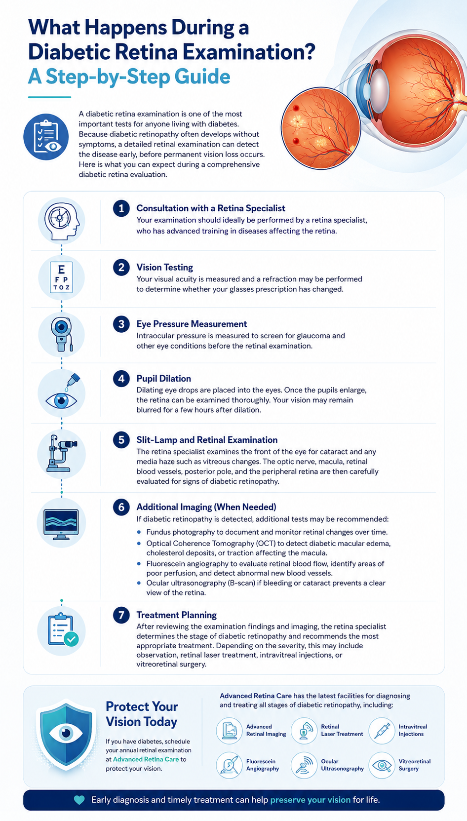
A diabetic retina examination is one of the most important tests for anyone living with diabetes. Because diabetic retinopathy often develops without symptoms, a detailed retinal examination can detect the disease early, before permanent vision loss occurs. Here is what you can expect during a comprehensive diabetic retina evaluation at Advanced Retina Care.
Step 1: Consultation with a Retina Specialist
Your examination begins with a detailed medical history consultation with Dr. Anina Abraham, Vitreo-Retina Specialist. You will discuss the duration of your diabetes, current HbA1c blood sugar levels, blood pressure, kidney health, past eye treatments, and any subtle visual symptoms you may have noticed, such as floaters or blurred reading vision.
Step 2: Visual Acuity Testing
Visual acuity is tested using a standardized eye chart for both distance and near vision. This measures your current visual sharpness and establishes a clinical baseline for future comparisons.
Step 3: Eye Pressure Measurement (Tonometry)
Intraocular pressure (IOP) is measured to screen for glaucoma and ensure eye pressure is normal prior to pupil dilation and further imaging.
Step 4: Pupil Dilation
Dilating eye drops are administered to enlarge your pupils. This allows the doctor to view the entire retina, macula, optic nerve, and peripheral blood vessels. Dilation typically takes 20–30 minutes and causes temporary mild blurring and light sensitivity lasting 3–4 hours.
Step 5: Slit-Lamp and Indirect Ophthalmoscopy
Using a specialized slit-lamp and high-magnification lenses, the retina specialist examines the posterior pole, macula, retinal vessels, and peripheral retina for microaneurysms, hemorrhages, exudates, swelling, or abnormal blood vessel growth.
Step 6: Specialized Diagnostic Imaging (OCT & FFA)
If diabetic retinopathy or macular edema is detected, state-of-the-art imaging may be performed:
- Optical Coherence Tomography (OCT): A non-invasive 3D cross-sectional scan measuring macular thickness and swelling down to the micron level.
- Fundus Photography: High-resolution digital color photos to document baseline retinal findings and monitor changes over time.
- Fundus Fluorescein Angiography (FFA): Dye-based imaging to highlight blocked capillaries, areas of poor blood flow (ischemia), and leaking abnormal blood vessels.
- Ocular Ultrasonography (B-Scan): Ultrasound imaging used if vitreous bleeding or cataract obscures a clear view of the retina.
Step 7: Personalised Treatment Planning
Based on diagnostic results, Dr. Anina Abraham explains the findings and creates a tailored management plan—whether it involves regular observation, retinal laser photocoagulation, intravitreal Anti-VEGF injections, or vitreoretinal surgery.